Radiation Therapy Eye Cancer: What You Need to Know
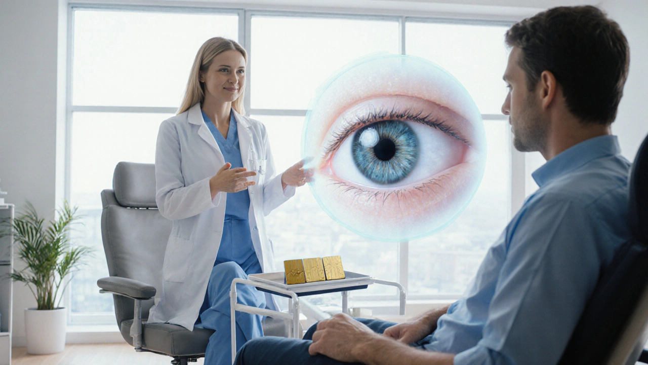
When dealing with radiation therapy eye cancer, a focused, high‑energy treatment aimed at destroying malignant eye cells while sparing surrounding tissue. Also called ocular radiation, it is a cornerstone for conditions like uveal melanoma, the most common primary eye cancer in adults, and other intra‑ocular malignancies. The approach blends physics, imaging, and surgical skill to target tumors that sit in delicate areas such as the retina or choroid.
Why choose radiation over surgery? In many cases the tumor’s location makes removal risky, and radiation can preserve vision better than enucleation. Radiation therapy eye cancer demands precise dose planning, which means a radiation oncologist works closely with an ophthalmic oncologist and a medical physicist to map the exact shape of the tumor. This collaboration ensures the beams hit the cancer cells while limiting exposure to the optic nerve and healthy retina.
Key Modalities: Brachytherapy vs. External Beam
Two main techniques dominate the field. Brachytherapy, also known as plaque therapy, involves attaching a tiny radioactive plaque to the sclera for several days. The plaque delivers a high dose right at the tumor site, reducing collateral damage. It’s ideal for medium‑size melanomas located near the front of the eye. In contrast, external beam radiation therapy (EBRT) uses machines like the proton beam or stereotactic radiosurgery to aim from outside the eye. EBRT is preferred for larger or posterior tumors that are hard to reach with a plaque.
Both modalities share a need for meticulous imaging—ultrasound, OCT, and MRI help define tumor dimensions to the millimeter. The treatment plan then translates those measurements into a 3‑D dose map, guiding the radiation physicist to shape the beam or plaque accordingly. This process embodies the semantic triple: "Radiation therapy eye cancer requires accurate imaging, which enables precise dose delivery".
Side effects are a reality, but modern techniques keep them manageable. Common issues include cataract formation, dry eye, and radiation retinopathy, which may appear months to years after treatment. Early detection through regular eye exams can mitigate vision loss. Patients are often counseled on protective measures like antioxidant supplements and UV‑blocking eyewear to support retinal health during recovery.
Follow‑up care doesn’t end when the plaque is removed or the beam is turned off. Long‑term monitoring includes visual acuity tests, retinal imaging, and sometimes fluorescein angiography to spot early signs of radiation‑induced damage. A multidisciplinary team—ophthalmic oncologist, radiation oncologist, medical physicist, and low‑vision specialist—coordinates this surveillance, ensuring any complication is caught early and treated promptly.
Understanding radiation therapy for eye cancer empowers you to ask the right questions, weigh benefits against risks, and stay proactive about eye health. Below you’ll find a curated set of articles that dive deeper into medication safety, cost‑effective drug sourcing, and related health topics—resources that can complement your treatment journey and help you make informed decisions about the broader aspects of care.